 ALERT
ALERT **Important for iLab Users**
The Help link in iLab opens an email to contact Support. To submit or track requests, visit: Submit and Track Support Requests


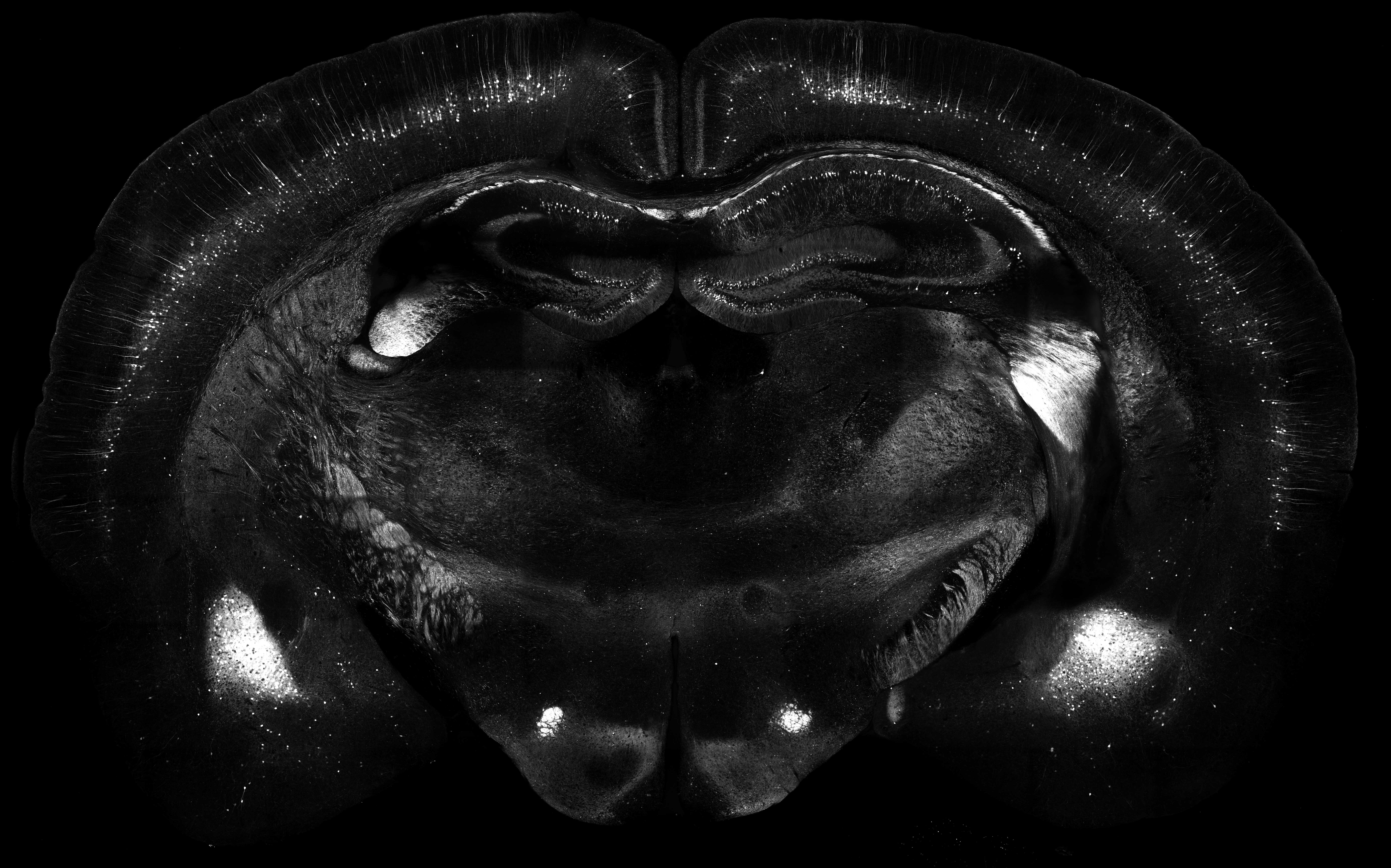
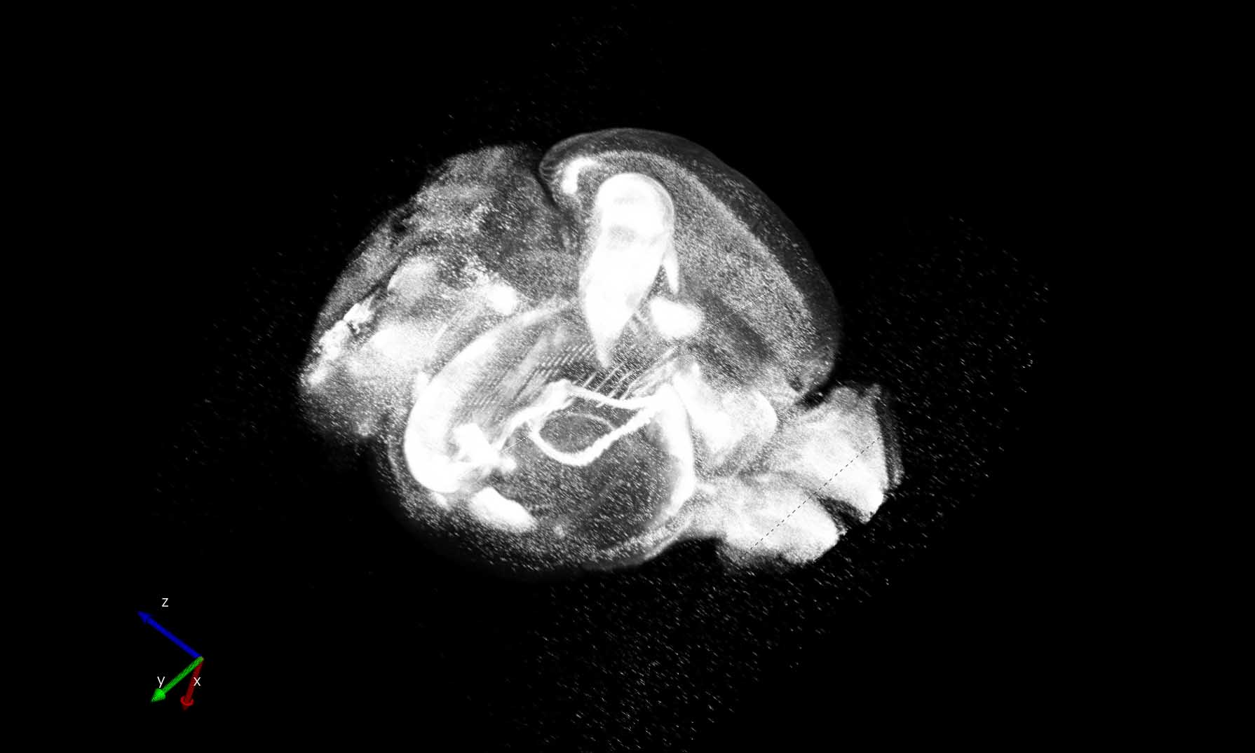
Serial two-photon images of a mouse model expressing YFP in subsets of neurons throughout the brain rendered in three dimensions. The brain was perfusion-fixed by standard methods and embedded in oxidized agarose in preparation for sectioning. The TissueCyte 1000 instrument (TissueVision, Inc.) automatically sectioned the entire volume of the brain at 100µm in the coronal plane and collected a mosaic of image tiles encompassing each section. The resulting images were corrected for uneven illumination and subjected to median filtering to reduce noise using custom MATLAB scripts. FIJI was used to stitch the processed tiles in the X-Y dimensions to create mosaic images of each tissue section. Neurolucida (Microbrightfield Bioscience) was used to render the images in three dimensions. A representative single coronal section (left panel) as well as the three-dimensional rendering of the whole brain (right panel) are shown.
The Whole Brain Microscopy Facility (WBMF), UT Southwestern Department of Neurology, was established in 2014 and is an Institutionally Supported Core (ISC). The facility is particularly well-suited to advance the study of traumatic brain injury as well as other neurological and psychiatric disorders by utilizing cutting-edge microscopy strategies to evaluate neuropathology across micro-, meso-, and macro-scales of inquiry. These techniques enable the tracing of neuronal projections from injured sites to their long-range targets, allowing crucial and previously unfeasible studies into the altered connections established by neurons following brain injury.
We provide access to several high-throughput microscopes for UT Southwestern and external researchers. WBMF houses two TissueVision TissueCyte 1000 instruments for the purpose of generating high-resolution, 3D renderings of entire rodent brains as well as four slide scanning microscopes (Hamamatsu Nanozoomer 2.0-HT, Hamamatsu Nanozoomer S60, Zeiss Axioscan.Z1 and Zeiss Axioscan 7) for the purpose of creating digital images of whole slides. We also offer cryosectioning, general histological staining such as H&E, cresyl violet, or Luxol Fast Blue, and immunostaining.
Denise Ramirez, PhD | Director
| Hours | Location |
|
Monday - Friday 8am - 5pm
|
UT Southwestern Medical Center Dept. of Neurology 5323 Harry Hines Blvd. Dallas, TX 75390-8813 |
| Name | Role | Phone | Location | |
|---|---|---|---|---|
| Denise Ramirez, PhD |
Director
|
214-648-0203
|
denise.ramirez@utsouthwestern.edu
|
NL10.125B
|
| Aleksandr Umanzor, BSc |
Research Technician II
|
Aleksandr.Umanzor@utsouthwestern.edu
|
NL10.110
|
|
| Li (Lily) Li, MD, MS |
Histology Technician
|
Li.Li@utsouthwestern.edu
|
NL10.110
|
|
| Ariana Nawaby, MS, BSc |
Computational Scientist
|
Ariana.Nawaby@utsouthwestern.edu
|
NL10.108
|
| Service list |
| ► TissueCyte Imaging (1) | |||
| Name | Description | Price | |
|---|---|---|---|
| rat brain, coronal sections, 0.875um/px resolution, 100um sections |
|
Inquire | |
| ► Training (1) | |||
| Name | Description | Price | |
| ilastik training |
hands on training to perform pixel classification in ilastik |
Inquire | |